The work was a controlled comparison, not a clinical stroke study.
Restricted-knee gait analysis using OpenSim.
This project investigated how mechanically restricting knee flexion changed lower-limb movement, joint moments and estimated muscle forces during walking.
ProblemUnderstand compensation caused by knee-flexion restriction
Approach3 conditions × 3 trials processed with OpenSim and MATLAB
Main resultAbout 10° lower peak right-knee flexion with bracing
My roleEqual contribution across collection, analysis, interpretation and reporting
Problem
What were we trying to find out?
Reduced knee flexion is one feature of post-stroke stiff-knee gait. Our group used a knee brace on one healthy participant to isolate this mechanical restriction and examine how the rest of the lower body responded.
Research question
How do baseline walking, a nominal 45° brace setting and a nominal 15° brace setting differ in joint angles, joint moments, ground-reaction forces and estimated muscle forces?
Marker trajectories were used to calculate joint motion.
Ground-reaction forces supported inverse dynamics.
Approach
How the data were collected and processed.
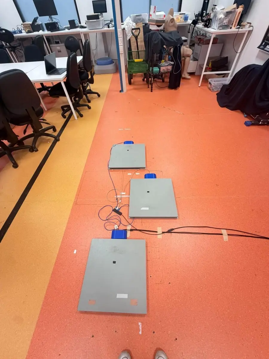
Three trials were recorded for each condition at a standardised cadence of 88 beats per minute. The model and trial outputs were then processed through a consistent workflow.
Fit the supplied musculoskeletal model to the participant.
Estimate joint angles from the marker trajectories.
Estimate net joint moments using motion and force data.
Estimate how muscle forces could produce the calculated motion.
MATLAB processing
- Imported the nine OpenSim outputs and matched equivalent variable names.
- Cropped each trial from the first force-plate heel strike to the final toe-off.
- Normalised each trial to 0–100% and interpolated it to 101 points.
- Averaged the three trials within each condition and produced comparison plots.
Result
What the analysis showed.
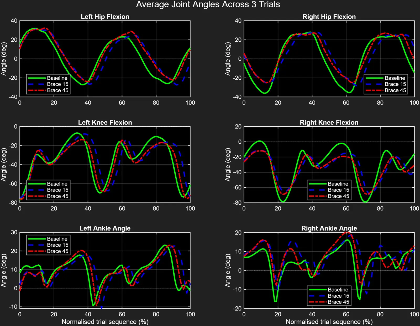
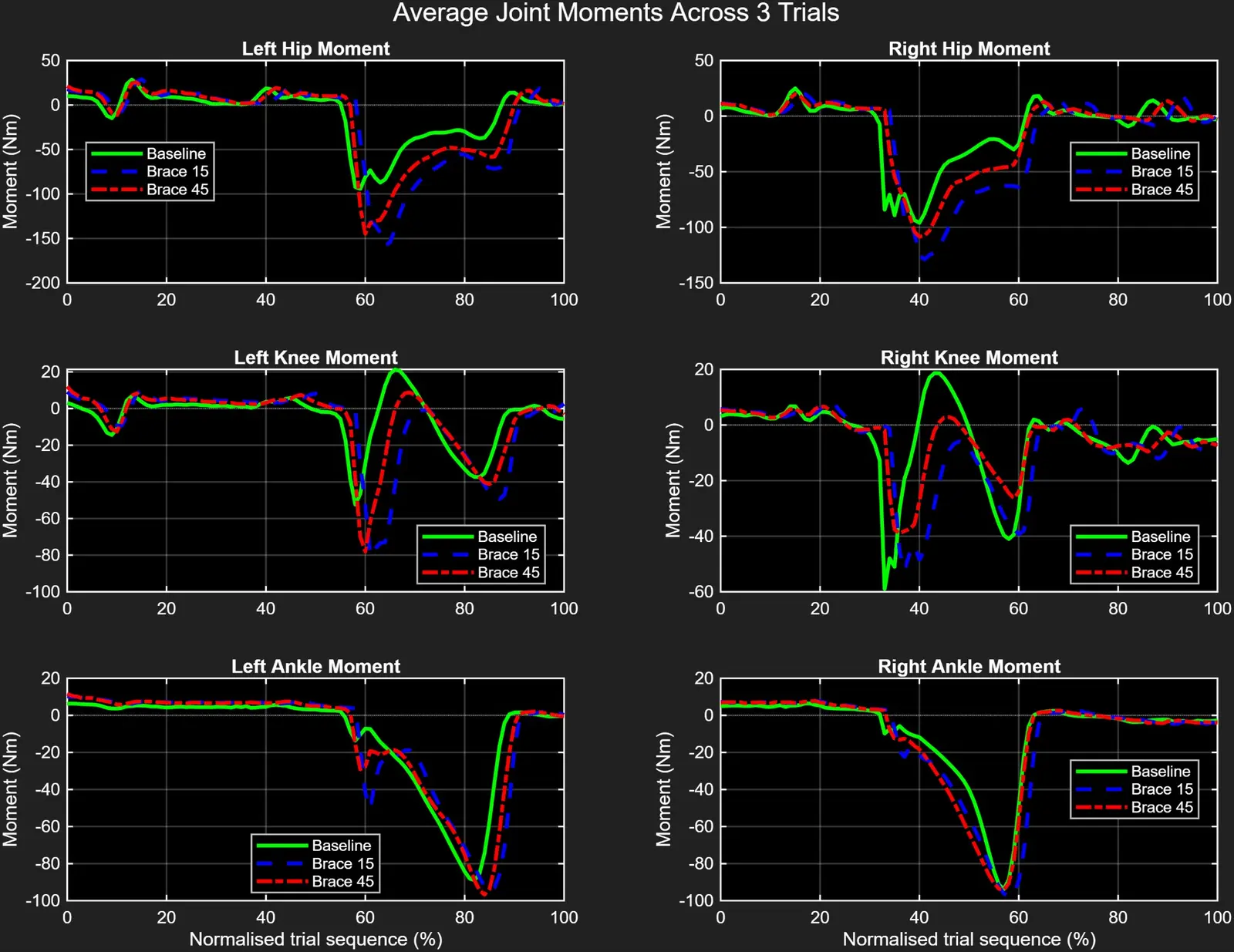
Both braced conditions reduced peak right-knee flexion from about 80° to about 70°. The main hip and knee moments also changed, and the model estimated lower right plantarflexor force with increased demand in parts of the opposite limb.
80° → 70°peak right-knee flexion magnitude
−95 → −130 N·mmain right-hip moment, baseline to Brace 15
3300 → 2200 Npeak right plantarflexor force
| Outcome | Baseline | Brace 15 | Brace 45 |
|---|---|---|---|
| Peak right-knee flexion | ≈80° | ≈70° | ≈70° |
| Main negative right-hip moment | ≈−95 N·m | ≈−130 N·m | ≈−110 N·m |
| First negative right-knee moment | ≈−60 N·m | ≈−50 N·m | ≈−40 N·m |
| Largest vertical GRF peak | ≈830 N | ≈780 N | ≈810 N |
Important result
The two brace conditions produced very similar knee motion and both exceeded their intended limits. The study showed adaptation to bracing, but it did not show that the 15° setting caused greater compensation than the 45° setting.
Reflection
What I learned.
The main lesson was that a model result is not the same as a direct measurement. OpenSim was useful for combining motion and force data, but every output depended on the model, marker placement, event timing and optimisation assumptions.
- I learned how scaling, inverse kinematics, inverse dynamics and Static Optimization are connected.
- I improved my MATLAB skills by processing repeated trials and keeping naming consistent across exported files.
- I learned to separate a useful trend from a claim that the data could not support.
- I improved my ability to present group results clearly and state the limits of the study.
Limitations
Where the work could have been stronger.
These were the main weaknesses of the work and the areas I would address if repeating the study.
- Brace validation: the achieved knee restriction was not checked independently, so the nominal settings were not actually produced in the model results.
- Small sample: one participant and three trials per condition limited generalisation and made variability difficult to assess.
- No variability bands: the final plots showed means without standard-deviation bands or individual-trial curves.
- Model validation: residuals, reserve actuators and EMG were not fully reported, so confidence in estimated muscle forces was limited.
- Clinical relevance: mechanical restriction in a healthy participant was only a simplified representation of stiff-knee gait after stroke.
What I would change
Measure the actual brace restriction, record walking speed, use gait-event-based comparison, add more participants and trials, show variability, and include EMG or other validation data.